shape
carat
color
clarity

Unexpected medical adventure

TooPatient

Super_Ideal_Rock
Premium
Joined
Sep 1, 2009
Messages
10,295
@ksinger definitely blood test first. They are lining blood test up with LP to make sure blood draw is just before LP.

Lucky too that one of the top MS neurologist people is with the hospital I am going through. Met him when we were dealing with DH's health issues.

Very glad my main coordinating doctor is working to find alternatives before approaching surgery. DH is also pushing to be sure all options are looked at rather than just jumping in.
 

yssie

Super_Ideal_Rock
Premium
Joined
Aug 14, 2009
Messages
27,263
I'm so sorry you have to go through this!!

I don't have any experience to share, but I will be thinking of you and hoping for a quick diagnosis and effective treatment... ::BIG HUGS::
 

missy

Super_Ideal_Rock
Premium
Joined
Jun 8, 2008
Messages
54,123
@TooPatient to answer your question I am sharing it here for you.
https://www.medscape.com/viewarticle/848225#vp_1

New diagnostic criteria have been introduced for neuromyelitis optica (NMO), which is now to be known as neuromyelitis optica spectrum disorder (NMOSD).

The new criteria were developed by an international consensus panel, headed by Dean Wingerchuk, MD, from the Mayo Clinic, Rochester, Minnesota, which reviewed the medical literature on NMOSD. The recommendations were published in the July 14 issue of Neurology.

They explain that NMOSD is an inflammatory disease of the central nervous system that can affect the optic nerves, brain stem, spinal cord, and brain. It can be mistaken for multiple sclerosis, but accurate diagnosis is essential because some drugs used for multiple sclerosis can worsen NMSOD.

NMOSD can cause a spectrum of symptoms, including visual loss, paralysis and episodes of persistent hiccups, nausea, and vomiting. A blood test to detect aquaporin-4 IgG antibodies (AQP4-IgG) is highly specific for NMOSD and facilitates the diagnosis, but some patients have the key features of NMOSD yet no detectable antibodies. The new criteria address both possibilities.

The new diagnostic criteria for NMOSD require at least one of the above core clinical characteristic with a positive result on an antibody test (cell based assay recommended).


If the antibody test result is not positive, then the patient must have at least two core clinical characteristics (one of which must be optic neuritis, acute myelitis with longitudinally extensive transverse myelitis lesions, or area postrema syndrome) and must also show additional MRI requirements.


The additional MRI requirements are as follows:

  • Acute optic neuritis: requires brain MRI showing (1) normal findings or only nonspecific white matter lesions or (2) optic nerve MRI with T2-hyperintense lesion or T1-weighted gadolinium enhancing lesion extending over 1/2 optic nerve length or involving optic chiasm

  • Acute myelitis: requires associated intramedullary MRI lesion extending over three contiguous segments or at least three contiguous segments of focal spinal cord atrophy in patients with history compatible with acute myelitis

  • Area postrema syndrome: requires associated dorsal medulla/area postrema lesions

  • Acute brainstem syndrome: requires associated periependymal brainstem lesions

In an accompanying editorial, Ilya Kister, MD, New York University School of Medicine, New York, and Friedemann Paul, MD, Charité University Medicine Berlin, Germany, point out that in just over a decade, "NMO has been transformed from an obscure, untreatable disorder…into a distinct nosologic entity with its own serologic marker and a wide range of as yet unproven, but universally deployed treatment options."


They attribute this "remarkable change of fortune" to the breakthrough discovery of AQP4-IgG antibody test, which they say is "exquisitely specific to NMO."


While the editorialists agree that the new diagnostic criteria will aid earlier identification of these patients and that it will be "highly desirable to identify and treat NMOSD after the first attack," there is still a risk for misdiagnosis with both false-positive and false-negative results.

They note that the use of cell-based assays for AQP4-IgG would help reduce the problem of misdiagnosis, but these are not widely available.


They add that "the best antidote to diagnostic error is a thorough familiarity with the clinical and paraclinical features that increase or decrease pretest probability of NMOSD" and point out that the new review document includes several useful examples in this regard.


These include the observation that onset-to-nadir time of less than 4 hours, or an inexorably progressive course, would be highly atypical for NMO myelitis. In addition, the presence of coexisting systemic lupus erythematosus, Sjögren syndrome, or myasthenia gravis increases confidence about NMOSD diagnosis, while extraneural sarcoidosis makes it less likely.


The International Panel for Neuromyelitis Optica Diagnosis was supported by the Guthy-Jackson Charitable Foundation. Dr Wingerchuk has received research support from Alexion, Terumo BCT, and the Guthy Jackson Charitable Foundation; receives financial compensation for participation on a relapse adjudication panel for MedImmune; and has served as a consultant to Alexion, MedImmune, and Chugai Pharmaceuticals. Other members of the panel receive royalties for a technology license related to a test for aquaporin-4 autoantibodies for diagnosis of neuromyelitis optica. Both editorialists are members of International Clinical Consortium of the Guthy-Jackson Charitable Foundation.


Neurology. 2015;85:177-189. Full text Editorial
"It's very important for clinicians to know about these updated criteria," said Dr Wingerchuk. "An accurate diagnosis is necessary for selecting the right therapy."

"In the past the previous criteria for a diagnosis of NMO required that the patient had both optic neuritis and transverse myelitis. And these are still the most common symptoms. But the new criteria allow other symptoms to be taken into account as well. These include postrema clinical syndrome characterized by intractable hiccups or nausea and vomiting, and other criteria involving the brainstem such as double vision or ataxia," he told Medscape Medical News.

"If a patient has any of these symptoms or an MRI scan with suggestive findings then an antibody test should be obtained. The new criteria allow for a diagnosis of NMOSD after a single attack if the antibody test is positive. It also allows the diagnosis to be given if the antibody test is negative under more stringent criteria. We believe more people will be captured after their first attack with these new criteria, which will lead to earlier and more accurate diagnosis."

The authors note the new consensus definition of NMOSD unifies traditional NMO and modern NMOSD definitions.

"It allows for NMOSD diagnosis in AQP4-IgG–seropositive patients with involvement of almost any CNS [central nervous system] region as well as in those with restricted involvement of a single region (eg, recurrent transverse myelitis)," they write.

For the first time, criteria allow for an NMOSD diagnosis in patients who have not experienced clinical involvement of optic nerves or spinal cord.

Core clinical characteristics of NMSOD are listed as:

  • Optic neuritis

  • Acute myelitis

  • Area postrema syndrome: episode of otherwise unexplained hiccups or nausea and vomiting

  • Acute brainstem syndrome

  • Symptomatic narcolepsy or acute diencephalic clinical syndrome with NMOSD-typical diencephalic MRI lesions

  • Symptomatic cerebral syndrome with NMOSD-typical brain lesions.
 

ksinger

Ideal_Rock
Premium
Joined
Jan 30, 2008
Messages
5,083
@ksinger definitely blood test first. They are lining blood test up with LP to make sure blood draw is just before LP.

Lucky too that one of the top MS neurologist people is with the hospital I am going through. Met him when we were dealing with DH's health issues.

Very glad my main coordinating doctor is working to find alternatives before approaching surgery. DH is also pushing to be sure all options are looked at rather than just jumping in.

Excellent! And so glad you have a MS specialist there. :)

Gotta fly! Will check in later.
 

diamondringlover

Ideal_Rock
Premium
Joined
Dec 12, 2006
Messages
4,410
my goodness, you have my thoughts and prayers!
 

PintoBean

Ideal_Rock
Premium
Joined
Jul 27, 2011
Messages
6,589
{{{hugs TP}}}
 

TooPatient

Super_Ideal_Rock
Premium
Joined
Sep 1, 2009
Messages
10,295
Had a thought... Going to ask nurse to ask doctor, but.... Why am I here?

LP is not until tomorrow morning. Nothing else between now and then. Why am I lying here in bed when I am stable on my feet? Not dizzy or anything.

Why can't I go home and come back for appointments?
 

Jambalaya

Ideal_Rock
Premium
Joined
Oct 2, 2014
Messages
4,784
Ask them. It's better to go home if it's safe. Less chance of getting an infection!
 

cflutist

Ideal_Rock
Premium
Joined
Jul 12, 2004
Messages
4,054
@TooPatient

Gosh, I am sorry that you are going through this stressful time.
Sending positive and caring thoughts your way, one step at a time, it sounds that you are at a top Neuro hospital and in good hands.

Yes, I underwent a pterional craniotomy to ressect a 3cm (walnut sized) meningioma 3 years ago. The incision was closed with 52 staples across my scalp. My bone flap is about 3.5cm by 4.5 cm, held together to my skull with 3 titanium plates, and 14 screws. Before this, we did meet with a Neuro Interventional Radiogist who advised that CyberKnife (targeted radiation) was not recommended due to the size of my tumor.

If you should proceed to surgery, I can answer recovery questions for you, but I am obviously not a doctor.

As to why you are in the hospital, my only guess is that they want to keep you under observation. Things related to Neuro can change very quickly, which is why I was awakened every hour on the hour post OP for Neuro checks.

Good luck to you.
 

TooPatient

Super_Ideal_Rock
Premium
Joined
Sep 1, 2009
Messages
10,295
Ask them. It's better to go home if it's safe. Less chance of getting an infection!

Besides... I would love to go pee without a nurse watching me!
 

TooPatient

Super_Ideal_Rock
Premium
Joined
Sep 1, 2009
Messages
10,295
@TooPatient

Gosh, I am sorry that you are going through this stressful time.
Sending positive and caring thoughts your way, one step at a time, it sounds that you are at a top Neuro hospital and in good hands.

Yes, I underwent a pterional craniotomy to ressect a 3cm (walnut sized) meningioma 3 years ago. The incision was closed with 52 staples across my scalp. My bone flap is about 3.5cm by 4.5 cm, held together to my skull with 3 titanium plates, and 14 screws. Before this, we did meet with a Neuro Interventional Radiogist who advised that CyberKnife (targeted radiation) was not recommended due to the size of my tumor.

If you should proceed to surgery, I can answer recovery questions for you, but I am obviously not a doctor.

As to why you are in the hospital, my only guess is that they want to keep you under observation. Things related to Neuro can change very quickly, which is why I was awakened every hour on the hour post OP for Neuro checks.

Good luck to you.

Thank you! I remembered you had something but couldn't recall the details.

They are only checking vitals every 4 hours and they have been steady. Squeeze fingers and stuff just once per day so far.
 

azstonie

Ideal_Rock
Premium
Joined
Jul 1, 2014
Messages
3,769
Hi @ksinger, I worked at the hospital, where we saw patients with that horrifying headache present while still admitted---but if the right peeps/schedule weren't in Interventional Radiology (IR) when the headache came on, or if the nurses don't know about blood patch, patients suffered terribly for nothing; that is why my advice was to get the internist/ordering physician to get in orders as a PRN so as not to have TP suffer needlessly post procedure. If IR knows going in (haha) that blood patch may be requested later, you tend to get it quicker in my experience.

LP in the clinic setting? NO THANK YOU! Hospital. Had one patient who, when the tech brought in the instrument tray, saw the needle and had a heart attack. Good thing he was at a hospital and not some clinic (hello Joan Rivers et alia).
 

cflutist

Ideal_Rock
Premium
Joined
Jul 12, 2004
Messages
4,054
I talked to a neurosurgeon at UW after my friend in ID was diagnosed with a glioblastoma after imaging at a visit to the ER. One thing he did mention to me is that friends/family of neurologists do NOT get yearly dental x-rays (or any other imaging of their heads unless a serious reason presents itself). He has been a neurologist for 30+ years and he feels there is an increase in tumors due to constant dental x-raying. I no longer allow my dentist to get imaging on a yearly basis ("But your insurance will cover it!").


This is from WebMD Archives:

Dental X-rays Linked to Brain Tumors

Annual X-rays May Expose Patients to Unnecessary Risk

By Salynn Boyles
This article is from the WebMD News Archive
This content has not been reviewed within the past year and may not represent WebMD's most up-to-date information.

To find the most current information, please enter your topic of interest into our search box.

FROM THE WEBMD ARCHIVES
April 10, 2012 -- Getting frequent dental X-rays appears to increase the risk for a commonly diagnosed brain tumor, a new study finds.

Exposure to ionizing radiation -- the kind found in X-rays -- is the biggest known environmental risk factor for largely non-malignant meningiomabrain tumors. Routine dental X-rays are among the most common sources of radiation for most healthy people in the U.S.

The new study suggests that performing frequent X-rays may expose patients to unnecessary risk.

"These findings should not prevent anyone from going to the dentist," says lead researcher and neurosurgeon Elizabeth B. Claus, MD, PhD, of Yale University School of Medicine and Boston's Brigham and Women's Hospital. "But it appears that a large percentage of patients receive annual X-rays instead of every two to three years, which is the recommendation for healthy adults."

Dental X-ray, Benign Brain Tumors
While the vast majority of meningiomas are non-malignant, they often grow to be very large and can cause a wide range of potentially serious symptoms, including vision and hearing loss, frequent headaches, memory loss, and even seizures.

They are the most frequently diagnosed brain tumors among adults in the United States, accounting for about a third of all primary brain and central nervous system tumors.
***************************************************************************

Another Viewpoint:

"The level of radiation used in a typical dental x-ray is extremely low. A full-mouth series of radiographs – which involves 18 separate pictures of the teeth – exposes an individual to roughly the same radiation risk as a plane passenger would absorb during a cross-country flight. At such levels, radiation is thought to have little or no effect on the risk for cancer.

Some studies have purported to show a link between dental x-rays and thyroid cancer and certain forms of brain cancer such as gliomas and meningiomas, but the results of such studies have been questioned because of a problem known as “recall bias.” Instead of showing a direct, physical connection between x-rays and cancer, these studies have relied on patients’ recollection of how many radiographs they’ve had during their lifetime.

Today, dental patients are exposed to even lower levels of radiation than in the past, as a result of digital x-ray technology, more precise radiation beams, and the addition of collars to x-ray shields placed over patients’ chests. When considering a dental x-ray, it’s important for patients to weigh the potential benefits – early and accurate diagnosis of oral and dental diseases – against the very minimal risks, says Jennifer Frustino, DDS, PhD, an oral medicine expert with Dana-Farber/Brigham and Women’s Cancer Center."

******************************************************

Cumulative exposure to radiation. That is what neurologists are concerned about here. If I don't have a toothache, I don't want risk and for some toothaches I want action taken so no need for imaging any way.

My friend in Neurology who first mentioned dental imaging and tumors to me, he was noticing it in his practice over time. Many more patients with brain tumors than he 'should' have been seeing.

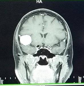
Thanks for your post. I had read this also and wondered if it was the cause of my meningioma seen here.

20180319_144950.jpg
 

ksinger

Ideal_Rock
Premium
Joined
Jan 30, 2008
Messages
5,083
Hi @ksinger, I worked at the hospital, where we saw patients with that horrifying headache present while still admitted---but if the right peeps/schedule weren't in Interventional Radiology (IR) when the headache came on, or if the nurses don't know about blood patch, patients suffered terribly for nothing; that is why my advice was to get the internist/ordering physician to get in orders as a PRN so as not to have TP suffer needlessly post procedure. If IR knows going in (haha) that blood patch may be requested later, you tend to get it quicker in my experience.

LP in the clinic setting? NO THANK YOU! Hospital. Had one patient who, when the tech brought in the instrument tray, saw the needle and had a heart attack. Good thing he was at a hospital and not some clinic (hello Joan Rivers et alia).

Interesting. Thanks for the insight. I'm just glad I didn't have any issues. As I said, 4 hours flat was boring, but I was all about boring versus the Headache From Hell.

And why does that highlighted statement not surprise me? :rolleyes:
 

kmarla

Brilliant_Rock
Premium
Joined
Feb 8, 2013
Messages
690
TooPatient, I’m so sorry to read this. I have to say my first thought was MS. My cousin has MS and ON is one of her symptoms. Whatever this turns out to be it sounds like you are receiving excellent care which is comforting. I am sending you strength and support and hope that you have the answers you need soon {{{hugs}}}.
 

Slickk

Ideal_Rock
Premium
Joined
Jan 3, 2013
Messages
5,006
Omgoodness how scary. No relevant advice, just lots of thoughts and hugs coming your way. I am so happy that you have a great team in a renowned hospital and a supportive dh. All of this will prove vital in getting to the bottom of this. ((Hugs))
 

azstonie

Ideal_Rock
Premium
Joined
Jul 1, 2014
Messages
3,769
@cflutist, that is a real mamajama on imaging :eek2::eek2::eek2:!!! You deserve a diamond of similar size :love: oh wait, you're the PSer who already does (counting carats of your beautiful jewels)!:mrgreen:

Toopatient, be sure to get your imaging and Rads reports emailed to you, so very soon you caratize them :lol::lol-2::mrgreen:
 

TooPatient

Super_Ideal_Rock
Premium
Joined
Sep 1, 2009
Messages
10,295
@cflutist, that is a real mamajama on imaging :eek2::eek2::eek2:!!! You deserve a diamond of similar size :love: oh wait, you're the PSer who already does (counting carats of your beautiful jewels)!:mrgreen:

Toopatient, be sure to get your imaging and Rads reports emailed to you, so very soon you caratize them :lol::lol-2::mrgreen:

Luckily (unlucky for caratizing...) mine are tiny.
 

TooPatient

Super_Ideal_Rock
Premium
Joined
Sep 1, 2009
Messages
10,295
Okay, so I am staying overnight.

The neurologist came in and is thinking more in line with MS or similar. He ordered addotaddit MRIs to be done overnight. These will show (hopefully) the chemical balance? This would help identify tumor or not. Also doing an atavan (so?) to rule out seizures (which would point toward tumor).

6:45am blood work
9:00am LP
12:15 neurological opthalmologist

Then we reevaluate and see where we go from there.
 

Bron357

Ideal_Rock
Premium
Joined
Jan 22, 2014
Messages
6,564
Sending healing hugs and wishing you a speedy recovery.
As awful as it is, take heart that these days they can do so many amazing things re brain surgery.
Hoping everything goes well for you and your family.
 

lyra

Ideal_Rock
Premium
Joined
Jul 13, 2007
Messages
5,249
Hi @ksinger, I worked at the hospital, where we saw patients with that horrifying headache present while still admitted---but if the right peeps/schedule weren't in Interventional Radiology (IR) when the headache came on, or if the nurses don't know about blood patch, patients suffered terribly for nothing; that is why my advice was to get the internist/ordering physician to get in orders as a PRN so as not to have TP suffer needlessly post procedure. If IR knows going in (haha) that blood patch may be requested later, you tend to get it quicker in my experience.

I had the headache after a LP gone wrong. It lasted a full week, and could have been avoided as you say, buy just laying flat for several hours. I couldn't even raise my head. Basically couldn't function. Definitely follow the correct protocol on this one. No one needs to go through that.
 

ksinger

Ideal_Rock
Premium
Joined
Jan 30, 2008
Messages
5,083
Okay, so I am staying overnight.

The neurologist came in and is thinking more in line with MS or similar. He ordered addotaddit MRIs to be done overnight. These will show (hopefully) the chemical balance? This would help identify tumor or not. Also doing an atavan (so?) to rule out seizures (which would point toward tumor).

6:45am blood work
9:00am LP
12:15 neurological opthalmologist

Then we reevaluate and see where we go from there.

So you're doing what type of MRIs overnight? (Having done 9 MRIs and a 10th scheduled for summer (sigh...) I'm just curious)
Are they going to do a spinal MRI? Ask for headphones tuned to your favorite radio station, cause that's a long one.

In a way, you're fortunate to present as you have. Eye problems means you get taken MUCH more seriously. You're going through quite the aggressive and compressed diagnosis process, which is good. So many people with neurological symptoms flail around for years trying to figure out what's wrong. It can be horribly frustrating a debilitating. My diagnosis was very fast for the world of neurological issues, it only took 2 months. You may get answers even sooner. I hope you do.

But in the privacy of your own head, now is the time for you to go back a few years and re-visit every single weird symptom you might have had, that was only a few weeks or months, that went away and you blew off. That time when your fingers on one hand kept missing the keys on your keyboard, or the recurring single intense itch on your foot, the phantom smell of electrical burning that was never there, or the numb patch on your back. Small things. Even the ones you think are silly to note, note them all. The doc will do an extensive dive into any potentially neurological symptoms you've EVER had. And they can be really subtle. And odd.

Here's hoping you get some sleep tonight in between all the other stuff. ;)2
 

DAF

Brilliant_Rock
Premium
Joined
Jan 7, 2012
Messages
778
TooPatient, so sorry about your troubles.

Although I am an Audiologist by profession, part of my work involves evaluation of stimulus conduction through various pathways, including the optic pathways, so I want to offer up some of my knowledge in hopes that it may direct you to the proper care.

When you said that they found lesions, I thought you meant plaque-like spots, much like ones one would see in Multiple Sclerosis. I was surprised to later read that they are considering biopsy, as if you had masses in your brain. OMG! Can you clarify?

One test to see where the visual signal is being held up is a test called Visual Evoked Response. You have to have some vision for this test to be completed, as it is affected by vision worse than 20/400. Anyway, you have surface electrodes at the back of your head above the inion (the part of the skull that juts out) while one eye is covered and the other watches a reversing checkerboard pattern. It's the change in pattern that registers at the back of the eye and causes the nerves to fire, sending the signal along the optic nerve, across the optic chiasm (the point at which what you see in, say, your left eye, crosses over to the right side of your brain and vice versa. In general, the signal crosses the optic chiasm at about 100 milliseconds, +/-. They may be able to localize the vision loss to anterior (in front of) or posterior (behind) to the optic chiasm. I've done this test for people with optic neuritis and as part of a diagnostic battery for diagnosing MS. This test looks for issues on the optic pathways, which may be totally unrelated to the lesions they've found.

Also, not part of my work, but from personal experience, is the thought of temporal arteritis, also called giant cell arteritis. It's autoimmune driven and can result in blindness. They have to confirm diagnosis by temporal artery biopsy. Preliminary blood work for this should evaluate ESR (erythrocyte sedimentation rate) and CRP ( C-Reactive Protein).

Hope this helps.
 

TooPatient

Super_Ideal_Rock
Premium
Joined
Sep 1, 2009
Messages
10,295
So you're doing what type of MRIs overnight? (Having done 9 MRIs and a 10th scheduled for summer (sigh...) I'm just curious)
Are they going to do a spinal MRI? Ask for headphones tuned to your favorite radio station, cause that's a long one.

In a way, you're fortunate to present as you have. Eye problems means you get taken MUCH more seriously. You're going through quite the aggressive and compressed diagnosis process, which is good. So many people with neurological symptoms flail around for years trying to figure out what's wrong. It can be horribly frustrating a debilitating. My diagnosis was very fast for the world of neurological issues, it only took 2 months. You may get answers even sooner. I hope you do.

But in the privacy of your own head, now is the time for you to go back a few years and re-visit every single weird symptom you might have had, that was only a few weeks or months, that went away and you blew off. That time when your fingers on one hand kept missing the keys on your keyboard, or the recurring single intense itch on your foot, the phantom smell of electrical burning that was never there, or the numb patch on your back. Small things. Even the ones you think are silly to note, note them all. The doc will do an extensive dive into any potentially neurological symptoms you've EVER had. And they can be really subtle. And odd.

Here's hoping you get some sleep tonight in between all the other stuff. ;)2

I don't remember the type ordered. It is a special thing that returns some ratio of chemicals from the spots they found.
I already had an MRI with and without contrast and some optical thing.
 

TooPatient

Super_Ideal_Rock
Premium
Joined
Sep 1, 2009
Messages
10,295
TooPatient, so sorry about your troubles.

Although I am an Audiologist by profession, part of my work involves evaluation of stimulus conduction through various pathways, including the optic pathways, so I want to offer up some of my knowledge in hopes that it may direct you to the proper care.

When you said that they found lesions, I thought you meant plaque-like spots, much like ones one would see in Multiple Sclerosis. I was surprised to later read that they are considering biopsy, as if you had masses in your brain. OMG! Can you clarify?

One test to see where the visual signal is being held up is a test called Visual Evoked Response. You have to have some vision for this test to be completed, as it is affected by vision worse than 20/400. Anyway, you have surface electrodes at the back of your head above the inion (the part of the skull that juts out) while one eye is covered and the other watches a reversing checkerboard pattern. It's the change in pattern that registers at the back of the eye and causes the nerves to fire, sending the signal along the optic nerve, across the optic chiasm (the point at which what you see in, say, your left eye, crosses over to the right side of your brain and vice versa. In general, the signal crosses the optic chiasm at about 100 milliseconds, +/-. They may be able to localize the vision loss to anterior (in front of) or posterior (behind) to the optic chiasm. I've done this test for people with optic neuritis and as part of a diagnostic battery for diagnosing MS. This test looks for issues on the optic pathways, which may be totally unrelated to the lesions they've found.

Also, not part of my work, but from personal experience, is the thought of temporal arteritis, also called giant cell arteritis. It's autoimmune driven and can result in blindness. They have to confirm diagnosis by temporal artery biopsy. Preliminary blood work for this should evaluate ESR (erythrocyte sedimentation rate) and CRP ( C-Reactive Protein).

Hope this helps.

Hi! They have used mass and lesion interchangeably to some degree. One specialist says looks like classic tumor. One says classic MS. (Not one and one, more like three and three...)

The neurologist today said lesion and like MS but not. So something in the inflammatory family.

I think the optical guy tomorrow is going to do a lot of the vision testing and looking at back thicknesses and stuff like that.

20/400 sounds horrible. I don't know what my eye falls at now, but I can't see a person standing two feet in front of me. Not even shapes, color, blob, etc. Just black nothingness when I look with the bad eye.
 

Calliecake

Ideal_Rock
Premium
Joined
Jun 7, 2014
Messages
9,237
Too Patient, I’m sorry you are dealing with all of this. It sounds like you are getting great care and have excellent doctors. Sending lots of dust and hugs your way. Callie
 

MissGotRocks

Super_Ideal_Rock
Premium
Joined
Jun 23, 2005
Messages
16,363
I am so sorry to hear of this TooPatient. Here's hoping that they have some definitive answers and treatment for you very soon. Please keep us posted as you can. Warm thoughts and hugs to you!
 

yennyfire

Ideal_Rock
Trade
Joined
Jun 6, 2010
Messages
6,872
I'm so very sorry TP! I will be keeping you in my prayers that all goes smoothly tomorrow and that you get some answers and relief asap!

So grateful to have so many knowledgeable folks here on PS!!
 

Dancing Fire

Super_Ideal_Rock
Premium
Joined
Apr 3, 2004
Messages
33,852
TP
Sorry to hear this news. I don't have any advice I can only offer you big hugs!
bighug5.gif
 
Be a part of the community Get 3 HCA Results
Top